Urinary Bladder Sonography Medicг Na Nemoci Studium Na 1 Lf Uk

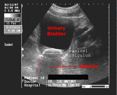
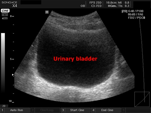
Urinary Bladder Sonography Medicг Na Nemoci Studium Na 1 о Filled bladder is visible with good assessment of its wall and we can also better see the organs located deep behind the bladder (uterus vagina in females). urinary bladder sonography | medicína, nemoci, studium na 1. Under normal circumstances it is located only in blood vessels (blood), in gallbladder and bile ducts (bile) and in urinary tract and bladder (urine). intra abdominal fluid sonography | medicína, nemoci, studium na 1.
Urinary Bladder Sonography Medicг Na Nemoci Studium Na 1 о Liver segments sonography | medicína, nemoci, studium na 1. lf uk. this is just an auxiliary text dedicated to liver functional anatomy. anatomic orientation in liver parenchyma has a big importance for sonography. liver segments are used for specific localisation of various pathologies in liver and for surgical liver resections. I have decided to work up some basic and practical information about abdominal sonography. this theme has been always very interesting to me, so i try to learn at least it‘s basics. there is a very unpleasant fact of insufficiency of czech written literature as well as practically non existing sources dedicated to abdominal ultrasound courses on the internet in both czech and english. the. Kalendář akcí. přednáškový večer chirurgické kliniky 2. lf uk a fnm. ocenění a oznámení. prof. brůha obdržel stříbrnou pamětní medaili univerzity karlovy. jedna zlatá a tři stříbrné medaile od vědecké rady uk pro osobnosti 1. lf uk. prof. ohad medalia obdržel čestnou vědeckou hodnost doctor honoris causa univerzity. Inferior vena cava sonography | medicína, nemoci, studium na 1. lf uk. the inferior vena cava (ivc) can be examined in transversal and sagittal direction – the same way as the abdominal aorta. it can be well displayed in the epigastrial area where we find ivc next to the aorta in front of vertebral body. except that we often visualize the.

Urinary Bladder Sonography Medicг Na Nemoci Studium Na 1 о Kalendář akcí. přednáškový večer chirurgické kliniky 2. lf uk a fnm. ocenění a oznámení. prof. brůha obdržel stříbrnou pamětní medaili univerzity karlovy. jedna zlatá a tři stříbrné medaile od vědecké rady uk pro osobnosti 1. lf uk. prof. ohad medalia obdržel čestnou vědeckou hodnost doctor honoris causa univerzity. Inferior vena cava sonography | medicína, nemoci, studium na 1. lf uk. the inferior vena cava (ivc) can be examined in transversal and sagittal direction – the same way as the abdominal aorta. it can be well displayed in the epigastrial area where we find ivc next to the aorta in front of vertebral body. except that we often visualize the. Pancreatic tail is better seen by a special maneuver put the probe classically into trasverse epigastrical projection, display the pancreas and then rotate the probe counterclockwise a bit (approximately 45 degrees). there are 4 main measurable sizes (widths) in pancreas: 1. pancreatic head – under 30mm. 2. pancreatic body – under 20mm. In abdominal sonography aorta may be sighted from upper epigastrium to iliac artery bifurcation. the view is possible in both sagittal and transverse section. aorta sonography | medicína, nemoci, studium na 1.

Comments are closed.