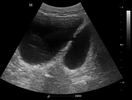
Urinary Bladder Diverticulum On Ultrasound And Ct Scan Case No 14

Urinary Bladder Diverticulum On Ultrasound And Ct Scan Case No 14 A urinary bladder diverticulum (plural: diverticula) is an outpouching from the bladder wall, whereby mucosa herniates through the bladder wall. it may be solitary or multiple in nature and can vary considerably in size. epidemiology. there are two peaks; one at 10 years and the other at 60 70 years 2. Urinary bladder diverticulum | radiology case | radiopaedia.org.

Urinary Bladder Diverticulum Radiology Reference Article Bladder diverticula are protrusions of the bladder urothelium and mucosa via muscle fibers of the bladder wall, the muscularis propria, which results in a thin walled structure connected to the bladder lumen and poorly empties during micturition. bladder diverticula occur either to congenital or acquired causes. they affect both adults and children. unlike the acquired adult form, in which. View ashesh ishwarlal ranchod's current disclosures. there are numerous causes of urinary bladder diverticula : primary (congenital or idiopathic) hutch diverticulum (in paraureteral region) secondary. bladder outlet obstruction. bladder neck stenosis. neurogenic bladder. posterior urethral valve. Introduction. bladder diverticula are defined as outpouchings of the urothelial lining through the muscularis layer of the bladder wall. they may result from congenital weakness of the bladder wall at the level of the ureterovesical junction (e.g. hutch’s diverticulum) or may be acquired as a result of increased intravesical pressure, typically in the context of lower urinary tract obstruction. The bladder diverticulum is an outpouching of the bladder wall (powell et al., 2009). in pseudodiverticula, only the mucous membrane of the bladder herniates; the diverticulum wall is without a muscle layer. in true diverticula, the outpouching consists of all bladder wall layers. ultrasound imaging of a small bladder diverticulum.

Diverticulum Of Bladder Ultrasound Introduction. bladder diverticula are defined as outpouchings of the urothelial lining through the muscularis layer of the bladder wall. they may result from congenital weakness of the bladder wall at the level of the ureterovesical junction (e.g. hutch’s diverticulum) or may be acquired as a result of increased intravesical pressure, typically in the context of lower urinary tract obstruction. The bladder diverticulum is an outpouching of the bladder wall (powell et al., 2009). in pseudodiverticula, only the mucous membrane of the bladder herniates; the diverticulum wall is without a muscle layer. in true diverticula, the outpouching consists of all bladder wall layers. ultrasound imaging of a small bladder diverticulum. A small urinary bladder diverticulum in a 78 year old man: (a) in the ca tomogram, against double opacification of the urinary bladder, with the patient lying faceup, a small diverticulum filled with the contrast medium is visible, and the diverticulum is located on the right rear wall of the urinary bladder (arrow). it is specially noted that. Highlights. an unusual case of multiple giant bladder diverticula required specialized surgery. the patient, 72, had severe symptoms due to a diverticulum compressing the ureter. treatment included catheterization, antibiotics, and prostate and diverticula resection. post surgery, the patient had improved urinary function without complications.

Urinary Bladder Diverticulum Radiology Reference Article A small urinary bladder diverticulum in a 78 year old man: (a) in the ca tomogram, against double opacification of the urinary bladder, with the patient lying faceup, a small diverticulum filled with the contrast medium is visible, and the diverticulum is located on the right rear wall of the urinary bladder (arrow). it is specially noted that. Highlights. an unusual case of multiple giant bladder diverticula required specialized surgery. the patient, 72, had severe symptoms due to a diverticulum compressing the ureter. treatment included catheterization, antibiotics, and prostate and diverticula resection. post surgery, the patient had improved urinary function without complications.

Diverticulum Of Bladder Ultrasound

Comments are closed.