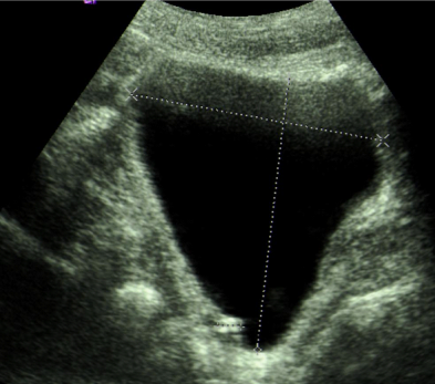
Ultrasound Picture Showing The Filling Defect In The Bladder

Ultrasound Picture Showing The Filling Defect In The Bladder Ultrasound is effective in the assessment of bladder diverticula. echo free outpouchings from the bladder are readily seen , and filling defects within a diverticulum such as stones or tumors also are visible. tumors within a diverticulum may present as soft tissue masses in the diverticulum or only as thickening of the diverticulum wall. A filling defect would be a dark spot somewhere in the esophagus amongst the white of the swallowed barium. the filling defect just means that something is preventing the white barium from covering that area. usually something that occupies space. another example would be a ct done to look at the bladder. in this case, contrast is given through.

Ultrasound Picture Showing The Filling Defect In The Bladder Computed tomography (ct) typically demonstrates an enhancing mass in the background of a urine filled bladder on contrast enhanced images and a filling defect of variable morphology in the background of a contrast filled urinary bladder in the excretory phase of imaging. calcifications have not been reported in bladder leiomyomas ( figure 68 2, a. Download scientific diagram | ultrasound picture showing the filling defect in the bladder. from publication: pheochromocytoma: a cause of anemia | patients with a pheochromocytoma usually present. The bladder mass exhibits variable degrees of postcontrast enhancement at the nephrographic phase, performed 100–120 s. after contrast medium injection. the tumoral mass shows washout and can be detected as a soft tissue filling defect in the contrast material filled hyperattenuating lumen on delayed phase of ct urography [28,29] (figure 4). (b) axial ct image obtained during the excretory phase and (c) sagittal excretory phase image show the same mass as a filling defect surrounded by excreted urine in the bladder. the most common method in determining nodal involvement by ct is size which is not reliable since small nodes may be metastatic and large nodes may be reactive.

Medcrave Online The bladder mass exhibits variable degrees of postcontrast enhancement at the nephrographic phase, performed 100–120 s. after contrast medium injection. the tumoral mass shows washout and can be detected as a soft tissue filling defect in the contrast material filled hyperattenuating lumen on delayed phase of ct urography [28,29] (figure 4). (b) axial ct image obtained during the excretory phase and (c) sagittal excretory phase image show the same mass as a filling defect surrounded by excreted urine in the bladder. the most common method in determining nodal involvement by ct is size which is not reliable since small nodes may be metastatic and large nodes may be reactive. A filling defect is a general term used to refer to any abnormality on an imaging study which disrupts the normal opacification (filling) of a cavity or lumen.the opacification maybe physiological, for example, bile in the gallbladder or blood in a dural venous sinus, or maybe due to the installation of contrast medium, for example within a vessel, as part of a ct angiogram, within the bladder. Filling of the bladder with contrast will reveal an irregular filling defect if the mass is indeed in the bladder. otherwise, cystography can show extrinsic compression of the bladder from an adjacent rms. however, this has been supplanted by sonography, ct, and mri .

Coronal Magnetic Resonance Image Of Bladder Demonstrating Filling A filling defect is a general term used to refer to any abnormality on an imaging study which disrupts the normal opacification (filling) of a cavity or lumen.the opacification maybe physiological, for example, bile in the gallbladder or blood in a dural venous sinus, or maybe due to the installation of contrast medium, for example within a vessel, as part of a ct angiogram, within the bladder. Filling of the bladder with contrast will reveal an irregular filling defect if the mass is indeed in the bladder. otherwise, cystography can show extrinsic compression of the bladder from an adjacent rms. however, this has been supplanted by sonography, ct, and mri .

Comments are closed.