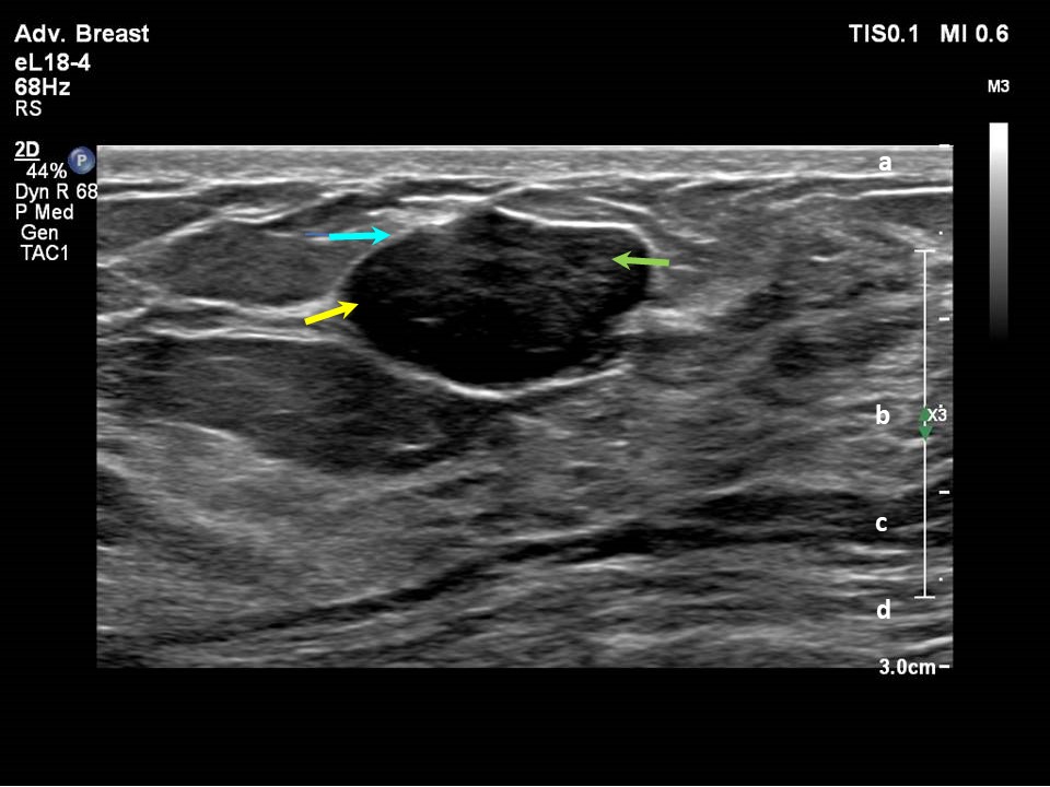
Ultrasound Of Bilateral Breasts A Anechoic Area With Echogenic Wall

Ultrasound Of Bilateral Breasts A Anechoic Area With Echogenic Wall Ultrasound of bilateral breasts. a anechoic area with echogenic wall, correlating with known breast implant. visualized implant wall demonstrates no disruption. b dimpling of the implant. Echogenicity is a descriptive term used to describe the picture that the reflected ultrasound waves form. each organ or body tissue has an expected echogenicity when it is not diseased. echogenicity can be used to compare an organ to its normal state or to another tissue. echogenicity can also be used to describe an abnormality on ultrasound.

Anechoic Ultrasound Solid masses of dense tissue are hypoechoic. hyperechoic. this term means "lots of echoes." these areas bounce back many sound waves. they appear as light gray on the ultrasound. hyperechoic. Here we see a normal ultrasound image of the breast. the upper grey layer is the skin. then there is a mixture of fat (dark or hypoechoic) and glandular tissue (light grey or hyperechoic). the striped layer posterior to the breast tissue is the pectoral muscle. posterior or deeper to the ribs there is a black area or posterior shadowing. Teaching points. at sonography, only 0.6 to 5.6% of breast masses are echogenic and the majority of these lesions are benign. approximately, 0.5% of malignant breast lesions appear hyperechoic on bus. echogenic lesions with worrisome features such as a spiculated margin, interval enlargement, or association with suspicious calcifications on. Imaging echogenic breast masses. the acr bi rads lexicon describes an echogenic breast mass on ultrasonography (us) as having an echogenicity greater than subcutaneous fat or equal to fibroglandular tissue. 1 an echogenic sonographic appearance is attributed histologically to fat, fibrous contents, vascular origin or high cellularity of lesions.

Anechoic Ultrasound Teaching points. at sonography, only 0.6 to 5.6% of breast masses are echogenic and the majority of these lesions are benign. approximately, 0.5% of malignant breast lesions appear hyperechoic on bus. echogenic lesions with worrisome features such as a spiculated margin, interval enlargement, or association with suspicious calcifications on. Imaging echogenic breast masses. the acr bi rads lexicon describes an echogenic breast mass on ultrasonography (us) as having an echogenicity greater than subcutaneous fat or equal to fibroglandular tissue. 1 an echogenic sonographic appearance is attributed histologically to fat, fibrous contents, vascular origin or high cellularity of lesions. Practice essentials. the first known clinical use of breast ultrasound (us) was reported in 1951 by wild and neal, [1] who used a mode sonography to describe the features of one benign and one malignant breast mass. several attempts were subsequently made to develop automated or multitransducer scanners to evaluate the whole breast, both to. Objective. breast ultrasound is helpful in the characterization of masses to differentiate benign from malignant disease. the internal echotexture of a mass is an important ultra sound feature in breast diagnostic workup. this article reviews the imaging and histopathology findings of benign and malignant hyperechoic masses to better recognize these conditions. conclusion. hyperechoic masses.

Ultrasound Images Of The Subareolar Areas Of Bilateral Breasts Practice essentials. the first known clinical use of breast ultrasound (us) was reported in 1951 by wild and neal, [1] who used a mode sonography to describe the features of one benign and one malignant breast mass. several attempts were subsequently made to develop automated or multitransducer scanners to evaluate the whole breast, both to. Objective. breast ultrasound is helpful in the characterization of masses to differentiate benign from malignant disease. the internal echotexture of a mass is an important ultra sound feature in breast diagnostic workup. this article reviews the imaging and histopathology findings of benign and malignant hyperechoic masses to better recognize these conditions. conclusion. hyperechoic masses.

Comments are closed.