Ultrasound Imaging Urinary Bladder Trabeculation 3d Ultrasound

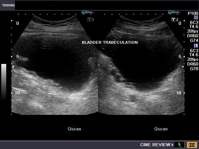
Ultrasound Imaging Urinary Bladder Trabeculation 3d Ultrasound Bladder outlet obstruction can arise from a number of conditions affecting the urethra and or bladder outlet but is most commonly encountered in elderly men due to prostate enlargement. clinical presentation. patients often present with difficulty in urination, retention, and urinary discomfort 2. Urinary bladder wall thickening is a common finding and its significance depends on whether the bladder is adequately distended. radiographic features ultrasound. in both adults and children, the wall may be considered thickened on ultrasound if it measures 6: >3 mm when distended (>25% expected volume*) >5 mm when non distended (<10% expected.

Ultrasound Imaging Urinary Bladder Trabeculation 3d Ultrasound However, 3d scanning demonstrated lattice like morphologic changes of the bladder wall which corresponded with the trabeculation and sacculation seen on cystoscopy. in that examination, bladder wall trabeculation, representing hypertrophied detrusor musculature, appears as interlaced muscular cords of different diameters. There is a long history of using two dimensional (2d) ultrasound imaging to estimate bladder volume [17,18] and more recently, three dimensional (3d) methods have become available . thus, the objective of this study was to compare three different ultrasound based methods of calculating bladder volume to identify the most accurate and precise. Background as trabeculated bladder wall is often referred to as a sign of chronically increased intravesical pressure, we investigated whether voiding cystourethrography (vcug) or sonography reliably predicts bladder trabeculation on later urethrocystoscopy.methods a total of 76 consecutive patients (2012–2017) with cystoscopically confirmed posterior urethral valves (puv) and pre endoscopy. The aim of our study was to assess the prognostic value of trabeculation of urinary bladder assessed by ultrasound as an non invasive diagnostic tool to diagnose the bladder outlet obstruction and prognostic factor to urinary retention and the need for surgery. out of 171 patients 120 were with spontaneous voiding and other 51 were presented.

Ultrasound Imaging Urinary Bladder Trabeculation 3d Ultrasound Background as trabeculated bladder wall is often referred to as a sign of chronically increased intravesical pressure, we investigated whether voiding cystourethrography (vcug) or sonography reliably predicts bladder trabeculation on later urethrocystoscopy.methods a total of 76 consecutive patients (2012–2017) with cystoscopically confirmed posterior urethral valves (puv) and pre endoscopy. The aim of our study was to assess the prognostic value of trabeculation of urinary bladder assessed by ultrasound as an non invasive diagnostic tool to diagnose the bladder outlet obstruction and prognostic factor to urinary retention and the need for surgery. out of 171 patients 120 were with spontaneous voiding and other 51 were presented. Female bladder outlet obstruction is uncommon. we report a case of bladder outlet obstruction secondary to urethral stenosis leading to bladder wall trabeculation. the patient presented at our clinic because of lower urinary tract symptoms including nocturia, urgency, bed wetting, hesitancy, straining to void, and incomplete emptying. Interest in using ultrasound technology for monitoring the urinary bladder through the lower abdomen has led to various wearable or portable ultrasound devices being proposed (supplementary note 2.

A Gallery Of High Resolution Ultrasound Color Doppler 3d Images Female bladder outlet obstruction is uncommon. we report a case of bladder outlet obstruction secondary to urethral stenosis leading to bladder wall trabeculation. the patient presented at our clinic because of lower urinary tract symptoms including nocturia, urgency, bed wetting, hesitancy, straining to void, and incomplete emptying. Interest in using ultrasound technology for monitoring the urinary bladder through the lower abdomen has led to various wearable or portable ultrasound devices being proposed (supplementary note 2.

Comments are closed.