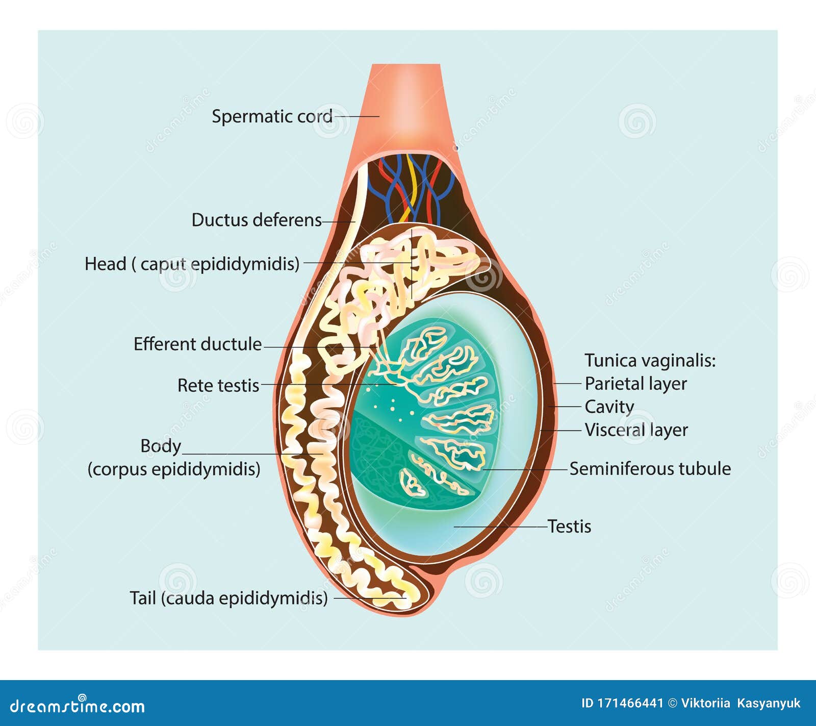
Testicle Epididymis Cross Section 3d Illustration Stock Illustratio

Testicle Epididymis Cross Section 3d Illustration Stock ођ Download testicle and epididymis, cross section stock illustration and explore similar illustrations at adobe stock. Find testicle and epididymis, cross section 3d illustration. testicle and epididymis, cross section royalty free stock images and million others at stockphotos.

Testicle Epididymis Cross Section 3d Illustration Stock ођ Find testicles testes illustration cross section testis stock images in hd and millions of other royalty free stock photos, 3d objects, illustrations and vectors in the shutterstock collection. thousands of new, high quality pictures added every day. 23.6 x 30.0 cm · 9.3 x 11.8 in (300dpi) request price add to basket add to board. credit. qa international science photo library. caption. the testicle is both an exocrine and endocrine gland. its exocrine activity consists in producing sperm cells, which are led from the testicle by a complex network of ducts (seminal tubes, epididymis, vas. Vector isolated illustration of human internal organs in male body. stomach, liver, intestine, bladder, lung, testicle, uterus, spine, pancreas, kidney, heart, bladder icon. donor medical poster. male and female reproductive system diagram. uterus and ovary, penis and testis in man and woman body. sexual organ anatomy concept. 4,327 testicle illustrations, drawings, stickers and clip art are available royalty free. find testicle stock images in hd and millions of other royalty free stock photos, illustrations and vectors in the shutterstock collection. thousands of new, high quality pictures added every day.

Testicle Epididymis Cross Section 3d Illustration Stock ођ Vector isolated illustration of human internal organs in male body. stomach, liver, intestine, bladder, lung, testicle, uterus, spine, pancreas, kidney, heart, bladder icon. donor medical poster. male and female reproductive system diagram. uterus and ovary, penis and testis in man and woman body. sexual organ anatomy concept. 4,327 testicle illustrations, drawings, stickers and clip art are available royalty free. find testicle stock images in hd and millions of other royalty free stock photos, illustrations and vectors in the shutterstock collection. thousands of new, high quality pictures added every day. The testis is both an exocrine (producing spermatozoa) and an endocrine (producing androgens) gland. this immature testis, cut in cross section, provides an overview of the testis proper, its enclosing tunics, and its location in the scrotum. also seen are the epididymis and the ductus deferens lying posterior to the testis. immature testis 10x. Caption. testicles anatomy. 1866 anatomical illustrations of the testes (testicles). fig. 1: frontal view of the testes (lower left and right) inside the scrotum, with a section through the penis (centre) and pubic hair at top. fig. 2: profile view showing a single testis (round, bottom right) and associated blood supply (red), epididymis (curled, lower right), vas deferens (vertical, white tube.

Illustration Of A Cross Section Of The Testis Epididymis Stock Vector The testis is both an exocrine (producing spermatozoa) and an endocrine (producing androgens) gland. this immature testis, cut in cross section, provides an overview of the testis proper, its enclosing tunics, and its location in the scrotum. also seen are the epididymis and the ductus deferens lying posterior to the testis. immature testis 10x. Caption. testicles anatomy. 1866 anatomical illustrations of the testes (testicles). fig. 1: frontal view of the testes (lower left and right) inside the scrotum, with a section through the penis (centre) and pubic hair at top. fig. 2: profile view showing a single testis (round, bottom right) and associated blood supply (red), epididymis (curled, lower right), vas deferens (vertical, white tube.

Comments are closed.