Structure Of Epithelial Tissue Free Biology Notes Rajus Biology

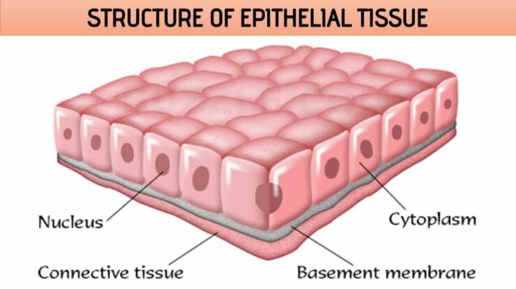
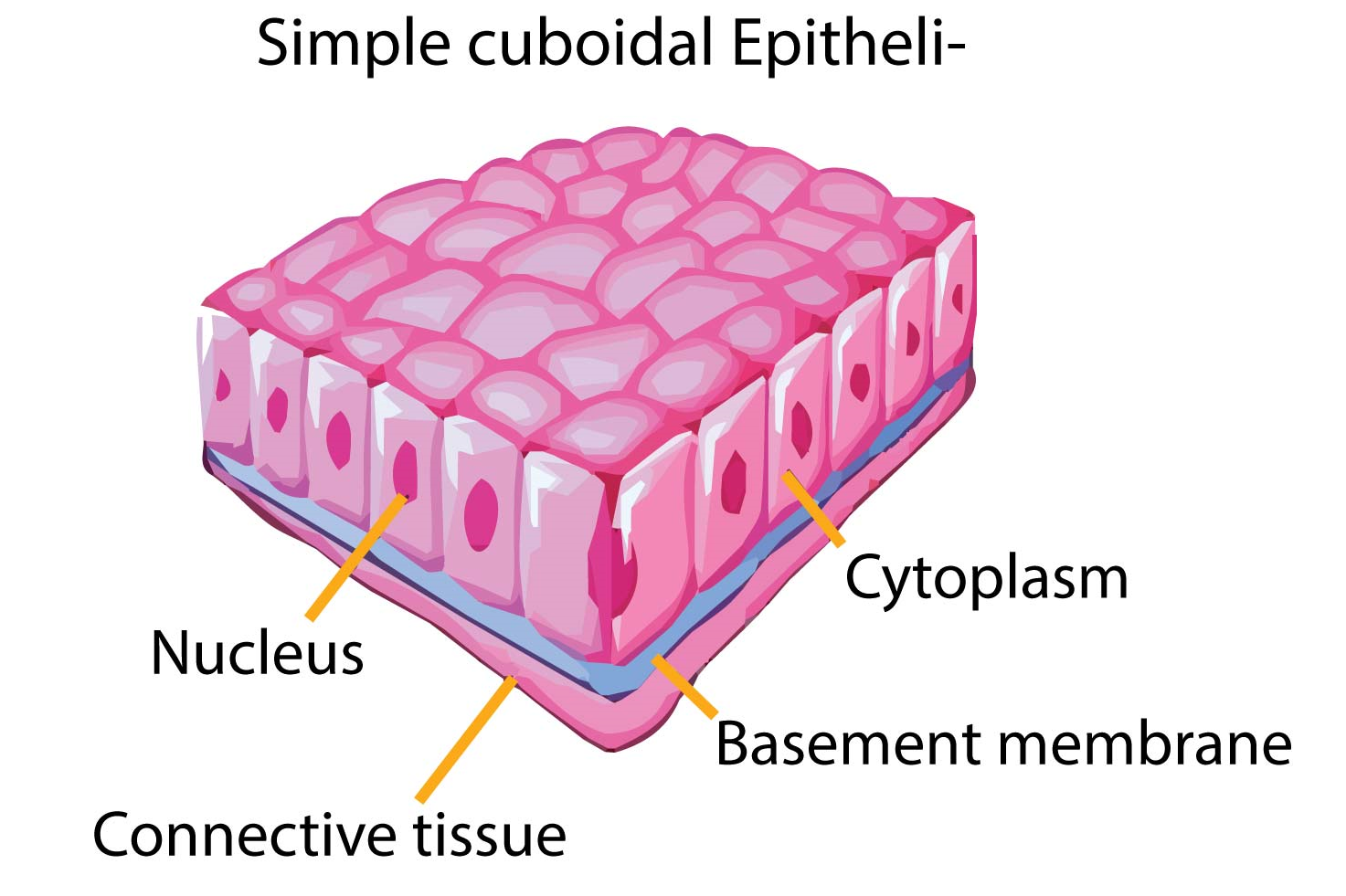
Structure Of Epithelial Tissue Free Biology Notes Rajus Biology One surface of epithelial tissue is attached to membrane, which consists of fibres and polysaccharides called basement membrane. basement membrane consist of 2 layers. basal lamina: made up of glycoprotein, which is secreted by epithelium cells. fibrous lamina: made up of collagen and reticular fibres are suspended in mucopolysaccharide. On the basis of number of cell layers it has 2 types: simple epithelium and componund epithelium. 1. simple epithelium tissues. composed of single layer of cells. functions as lining for body cavities, ducts and tubes. on the bssis of shape structural modifications of cells. (a) simple squamous epithelial tissue.

Epithelial Tissue Structure Types And Function With Diagram Free This article we will discuss about plant tissue system notes: epidermal tissue system , ground tissue system & vascular tissue system. several tissues may collectively perform the same function. a collection of tissue performing the same function is known as tissue system. on the basis of location, position and structure, sachs categorised. Epithelial tissue is avascular, relying on the blood vessels of the adjacent connective tissue to bring nutrients and remove wastes. the exchange of substances between epithelial tissue and connective tissue occurs by diffusion. innervated. epithelial tissue is innervated; that is, it has its own nerve supply. Epithelial cells form membranes. the epithelial membrane consists of a layer of epithelial tissue and has underlying connective tissue. there are two types of epithelial membranes, mucous membrane and serous membrane. mucous membrane: it is also known as mucosa. there are goblet cells present, which secrete mucus. A tissue is a group of cells having a common origin, similar structure and function and held together by a cementing substance. example: connective tissue. different types of tissues working together and contributing to specific functions inside the body constitute an organ. example: stomach.

Epithelial Tissue Structure Types And Function With Diagram Free Epithelial cells form membranes. the epithelial membrane consists of a layer of epithelial tissue and has underlying connective tissue. there are two types of epithelial membranes, mucous membrane and serous membrane. mucous membrane: it is also known as mucosa. there are goblet cells present, which secrete mucus. A tissue is a group of cells having a common origin, similar structure and function and held together by a cementing substance. example: connective tissue. different types of tissues working together and contributing to specific functions inside the body constitute an organ. example: stomach. The cell membrane is a remarkable structure that has properties of a solid and a liquid. side by side phospholipids arranged in a bilayer (called a lipid bilayer). the solid part (the “mosaic”) is the variety of proteins etc. embedded in the bilayer. each phospholipid has a hydrophobic tail and a hydrophylic head. Cell specialisation (1.1.3) cells specialise by undergoing differentiation: a process that involves the cell gaining new sub cellular structures in order for it to be suited to its role. cells can either differentiate once early on or have the ability to differentiate their whole life (these are called stem cells).

Epithelial Cell Diagram Labeled The cell membrane is a remarkable structure that has properties of a solid and a liquid. side by side phospholipids arranged in a bilayer (called a lipid bilayer). the solid part (the “mosaic”) is the variety of proteins etc. embedded in the bilayer. each phospholipid has a hydrophobic tail and a hydrophylic head. Cell specialisation (1.1.3) cells specialise by undergoing differentiation: a process that involves the cell gaining new sub cellular structures in order for it to be suited to its role. cells can either differentiate once early on or have the ability to differentiate their whole life (these are called stem cells).

Comments are closed.