Pelvic Ultrasound Transverse Anatomy

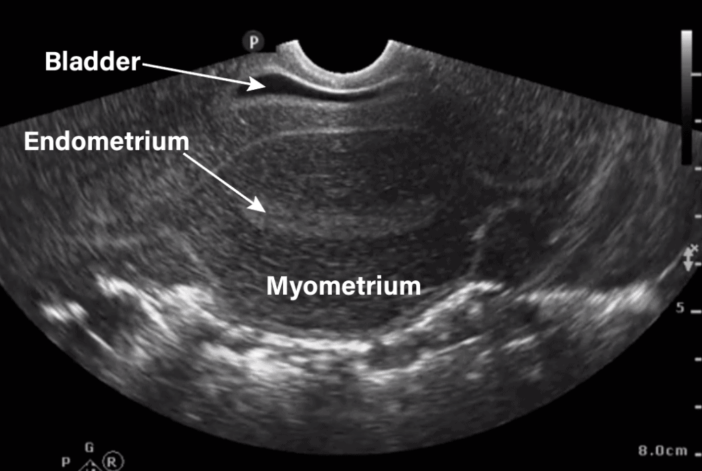
Transvaginal Pelvic Ultrasound Transverse And Sagitta Vrogue Co Step 2: obtain transverse view of uterus. rotate the probe 90 degrees counterclockwise with the indicator towards the patient’s right. obtain a good view of the fundus of the uterus as well as the endometrium and myometrium. tilt fan your probe through the entire uterus in the transverse view. The normal sonographic anatomy described in this chapter reflects a combined transabdominal and transvaginal approach and is weighted, as in clinical practice, to emphasize the positive attributes of both tvs and tas. fig 26 1. diagrammatic representation of the transvaginal ultrasound examination.

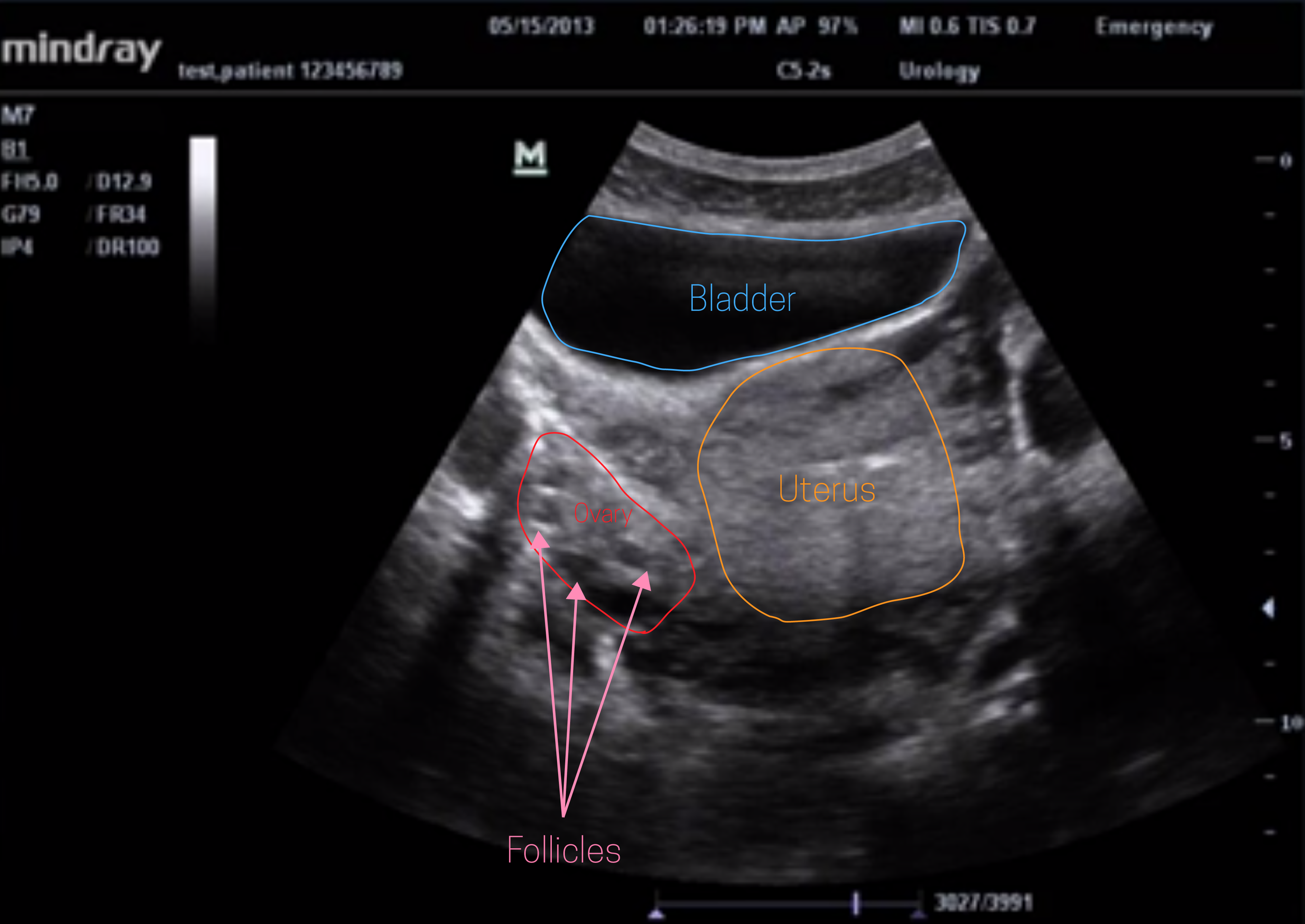
Ultrasound Leadership Academy The Basics Of Pelvic Transabdominal Anatomy and physiology. uterus cervix. the midline and parasagittal views are utilized to demonstrate the uterus from the fundus to cervix. the addition of transverse views allow for a complete assessment of size and contour, and the anatomy can be evaluated for abnormalities such as masses, typically benign leiomyomas. A transvaginal ultrasound offers an invaluable avenue for imaging the female pelvic anatomy. it augments transabdominal ultrasound for a more complete evaluation of the ovaries, adnexa, uterus, cervix, and surrounding pelvic regions. typically, an ultrasound technician will conduct the scanning and take images of the normal structures, with. Sweep the uterus in a transverse view, rotating the transducer 90° counterclockwise, such that the right side of the uterus is viewed on the left side of the ultrasound monitor. rotating the transducer in the counterclockwise manner maintains the same orientation of the pelvic anatomy and the image on the video monitor. Explore pulmonary hypertension using our cases which incorporate a virtual manikin for auscultation. use case studies to learn about abdominal sounds and their significance. cases include text, audio and assessment exercises. these exercises supplement our heart and lung sound courses and reference guides by helping users build listening.

Comments are closed.