Diagnosing Strokes With Imaging Ct Mri And Angiography Nclex Rn

Diagnosing Strokes With Imaging Ct Mri And Angiography Nclex Rn Visit us ( khanacademy.org science healthcare and medicine) for health and medicine content or ( khanacademy.org test prep mcat) for mcat. What sort of imaging can be done to diagnose stroke, and what do you actually see on these scans? have a look see!head on over to khanacademy.org or the khan.

Diagnosing Strokes With Imaging Ct Mri And Angiography Nclex Rn Stroke or cerebrovascular accident (cva) is an acute central nervous system (cns) injury and one of the leading causes of death in the developed world. estimates are that the incidence of stroke is 795000 each year, which causes 140000 deaths annually. based on the center for disease control and prevention (cdc) report, stroke has moved from third place in 2007 to the fifth leading cause of. Imaging of the acute stroke patient can be accomplished quickly and noninvasively with cta and mra. for occlusions of the major vessels at the skull base, these modalities are almost as accurate as dsa (loe: a). 4. imaging of chronic stenoses and occlusions can best be accomplished by ce mra, cta, and dsa. Comparison of ct and mri in stroke imaging. ct is widely used to initially evaluate patients with acute stroke symptoms within 24 h of stroke onset. in ct generated images, blood products appear as distinct hyperintense lesions, which can be identified in the hyperacute phase (between 0 and 6 h) of haemorrhagic stroke. Acute ischemic stroke is common and often treatable. imaging by computed tomography (ct) and mri is valuable for stroke treatment, in addition, for diagnosis, and for identification of the pathogenesis. but how they are used also depends on practical considerations. the approach to imaging the patient with acute stroke used at the massachusetts.

Khan Academy Diagnosing Strokes With Imaging Ct Mri And Comparison of ct and mri in stroke imaging. ct is widely used to initially evaluate patients with acute stroke symptoms within 24 h of stroke onset. in ct generated images, blood products appear as distinct hyperintense lesions, which can be identified in the hyperacute phase (between 0 and 6 h) of haemorrhagic stroke. Acute ischemic stroke is common and often treatable. imaging by computed tomography (ct) and mri is valuable for stroke treatment, in addition, for diagnosis, and for identification of the pathogenesis. but how they are used also depends on practical considerations. the approach to imaging the patient with acute stroke used at the massachusetts. Background: the relative value of computed tomography (ct) and magnetic resonance imaging (mri) in acute ischemic stroke (ais) is debated. in may 2018, our center transitioned from using ct to mri as first line imaging for ais. this retrospective study aims to assess the effects of this paradigm change on diagnosis and disability outcomes. methods: we compared all consecutive patients with. Introduction. neuroimaging in the evaluation of acute stroke is used to differentiate hemorrhage from ischemic stroke, to assess the degree of brain injury, and to identify the vascular lesion responsible for the stroke. multimodal computed tomography (ct) and magnetic resonance imaging (mri), including perfusion imaging, can distinguish.

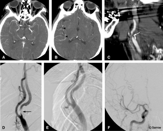
Ct Angiography And Stroke Barrow Neurological Institute Background: the relative value of computed tomography (ct) and magnetic resonance imaging (mri) in acute ischemic stroke (ais) is debated. in may 2018, our center transitioned from using ct to mri as first line imaging for ais. this retrospective study aims to assess the effects of this paradigm change on diagnosis and disability outcomes. methods: we compared all consecutive patients with. Introduction. neuroimaging in the evaluation of acute stroke is used to differentiate hemorrhage from ischemic stroke, to assess the degree of brain injury, and to identify the vascular lesion responsible for the stroke. multimodal computed tomography (ct) and magnetic resonance imaging (mri), including perfusion imaging, can distinguish.

Diagnosing Strokes By History And Physical Exam Nclex Rn Khan

Comments are closed.