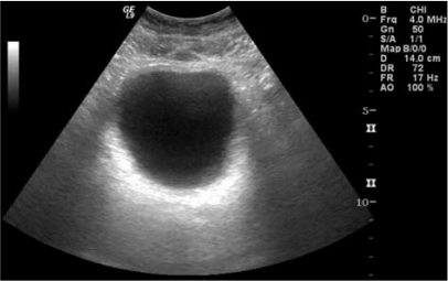
Bladder Ultrasonographic Finding Shows Mild And Diffuse Open I

Bladder Ultrasonographic Finding Shows Mild And Diffuse Open I Posted by sade96 @sade96, jan 30, 2023. hello, i just got my results back from my kidney and bladder, ultrasound. i’m the report is said i have mild thickening of my bladder wall 5 mm. i void at night 5 7 it’s so stressful. i also have hyperthyroidism and wondering if this is causing my bladder to thicken ?. Urinary bladder wall thickening is a common finding and its significance depends on whether the bladder is adequately distended. radiographic features. ultrasound. in both adults and children, the wall may be considered thickened on ultrasound if it measures 6: >3 mm when distended (>25% expected volume*).

Ultrasonographic View Of The Bladder In 2 Different Images The Image Bladder outlet obstruction. bladder outlet obstruction (boo) is a blockage at the base of the bladder where it empties into the urethra. for men, an enlarged prostate or prostate cancer can result. Diffuse bladder wall thickening is a common finding on ct but one which often does not indicate a specific diagnosis. diffuse bladder wall thickening is probably most commonly seen when the bladder is incompletely filled with urine. the bladder has to be fully distended with urine to say that the bladder wall thickening represents an abnormality. The thickness of the bladder will vary according to its distension. in a normal bladder, the wall decreases in thickness until it is half full (200 250 ml), and then stays more or less constant. the range of thickness is 3 5 mm, but with a full bladder it should be smooth and measure <3 mm. chronic diffuse thickening of the bladder is most. Ultrasonography (us) is the method of choice for the diagnosis of bladder disease. it is superior to other imaging techniques, such as urography and cystography, in depicting certain structures and abnormalities. us examination of the bladder should include a study of the ureterovesical junction and the structures round the vesical neck. the examination technique may be transabdominal.

Comments are closed.