Bladder Ct Scan

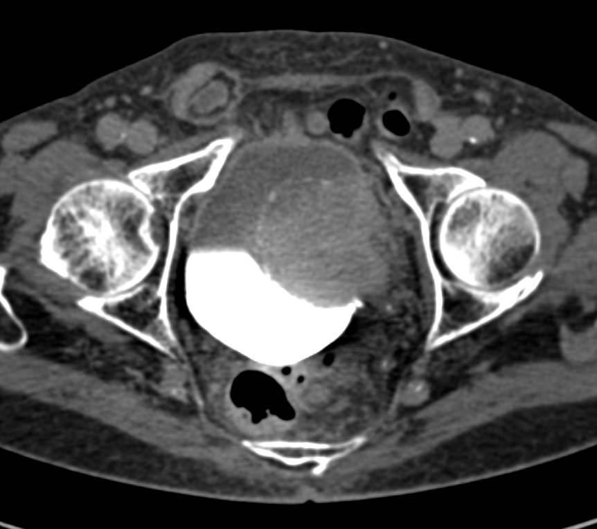
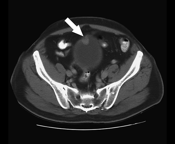
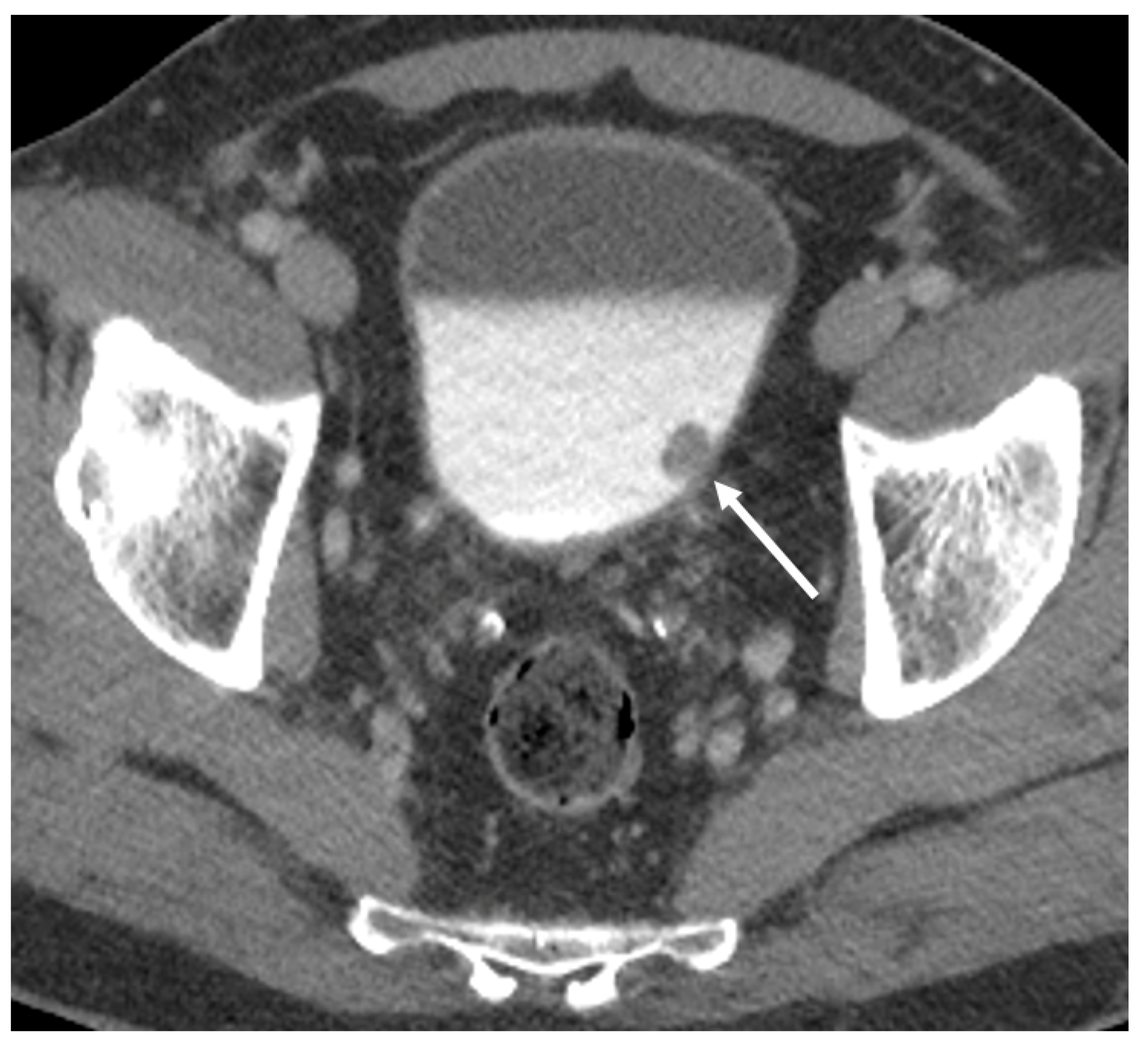
Bladder Cancer With Ct Urography Genitourinary Case Studies Ctisus A computerized tomography (ct) urogram is an imaging exam used to evaluate the urinary tract. the urinary tract includes the kidneys, bladder and the tubes (ureters) that carry urine from the kidneys to the bladder. a ct urogram uses x rays to generate multiple images of a slice of the area in your body being studied, including bones, soft. Ct scans are imaging tests that can detect and monitor bladder cancer with high accuracy. learn how they work, what to expect, and what other tests are available.

Detecting Bladder Cancer With A Ct Scan Ultrasound Or Mri Cxbladder A computed tomography (ct) urogram is a useful diagnostic tool for detecting conditions that affect your urinary system. it uses a series of x rays and a computer to produce three dimensional images of your soft tissues and bones. ct urograms are painless and have minor risks to your overall health. most people get their results in a few days. A ct urogram is a test that uses a ct scan and a special dye or contrast medium. it allows doctors to diagnose problems such as kidney and bladder stones, certain cancers, and structural. A ct scan of the abdomen and pelvis is used to check the urinary system for any tumours or blockages. it is also used to check if bladder cancer has spread to lymph nodes, the liver or other organs and tissue around the bladder. a ct scan of the chest may be used to check if bladder cancer has spread to the lungs. After confirming that you have bladder cancer, your doctor may recommend additional tests to determine whether your cancer has spread to your lymph nodes or to other areas of your body. tests may include: ct scan; magnetic resonance imaging (mri) positron emission tomography (pet) bone scan; chest x ray.

Diagnostics Free Full Text The Role Of Imaging In Bladder Cancer A ct scan of the abdomen and pelvis is used to check the urinary system for any tumours or blockages. it is also used to check if bladder cancer has spread to lymph nodes, the liver or other organs and tissue around the bladder. a ct scan of the chest may be used to check if bladder cancer has spread to the lungs. After confirming that you have bladder cancer, your doctor may recommend additional tests to determine whether your cancer has spread to your lymph nodes or to other areas of your body. tests may include: ct scan; magnetic resonance imaging (mri) positron emission tomography (pet) bone scan; chest x ray. The ct urogram is a radiological test to explore possible reasons for blood in the urine or other symptoms. this specialized scan uses intravenous (iv) contrast (a substance used to enhance the visibility of internal structures in x ray based imaging). a ct urogram examines the upper urinary tract (kidneys and ureters) in detail. Ct scans. a ct scan combines x rays with computer technology to create three dimensional (3 d) images. these scans can show stones in the urinary tract, as well as obstructions, infections, cysts, tumors, and traumatic injuries. imaging for urinary stone disease can be done with low or ultra low dose ct scans. radionuclide scans.

Tcc Bladder Radiology At St Vincent S University Hospital The ct urogram is a radiological test to explore possible reasons for blood in the urine or other symptoms. this specialized scan uses intravenous (iv) contrast (a substance used to enhance the visibility of internal structures in x ray based imaging). a ct urogram examines the upper urinary tract (kidneys and ureters) in detail. Ct scans. a ct scan combines x rays with computer technology to create three dimensional (3 d) images. these scans can show stones in the urinary tract, as well as obstructions, infections, cysts, tumors, and traumatic injuries. imaging for urinary stone disease can be done with low or ultra low dose ct scans. radionuclide scans.

Bladder Ct Scan

Comments are closed.