Bacterial Colony Morphologies вђ Pathelective
.jpeg)
Bacterial Colony Morphologies вђ Pathelective This is one of the first topics covered in the pathelective clinical microbiology module, so if you are interested, check out that course for more info. remember: the bacterial colony morphology might change depending on what agar it is on. for instance, proteus mirabilis is a motile, swarming bacterium, so when grown on blood agar, it can be. Welcome back to #micromeded. this week, i put together a simple figure describing the bacterial cell morphologies. this is a very important part of the microscopic examination of bacteria which, along with the gram stain, can sometimes give the clinician all they need to know to treat the patient (this is becoming less true as antimicrobial.

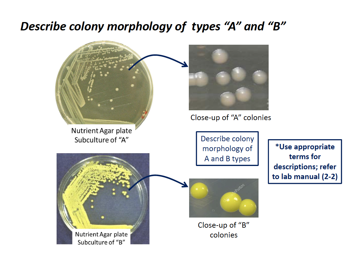
Bacterial Colony Morphology Chart This page titled 8: bacterial colony morphology is shared under a cc by nc sa 4.0 license and was authored, remixed, and or curated by jackie reynolds. bacteria grow on solid media as colonies. a colony is defined as a visible mass of microorganisms all originating from a single mother cell, therefore a colony constitutes a clone of bacteria. Bacteria grow on solid media as colonies. a colony is defined as a visible mass of microorganisms originating from a single mother cell. key features of these bacterial colonies serve as important criteria for their identification. characteristics of bacterial colonies. colony morphology can sometimes be useful in bacterial identification. Since morphology is influenced by medium type and growth conditions, care should be taken to record these parameters. good determination of colony morphology is predicated on good streak technique because it requires good separation of colonies. smibert and krieg (5) proposed the following protocol: 1. measure the colony diameter in millimeters. 2. The "bump" on an umbonate colony is called an umbo. 4. margin. erose is synonymous with serrated. 5. surface. surface can be smooth, glistening, rough, wrinkled, dry, powdery, moist, mucoid (forming large moist sticky colonies, brittle, viscous (difficult to remove from loop), butyrous (buttery). 6.

Bacterial Colony Morphologies Pathelective Vrogue Co Since morphology is influenced by medium type and growth conditions, care should be taken to record these parameters. good determination of colony morphology is predicated on good streak technique because it requires good separation of colonies. smibert and krieg (5) proposed the following protocol: 1. measure the colony diameter in millimeters. 2. The "bump" on an umbonate colony is called an umbo. 4. margin. erose is synonymous with serrated. 5. surface. surface can be smooth, glistening, rough, wrinkled, dry, powdery, moist, mucoid (forming large moist sticky colonies, brittle, viscous (difficult to remove from loop), butyrous (buttery). 6. The morphology of bacterial colony pattern is strongly dependent on the bacterial species and intercellular communication 26,27,28. in some cases, the bacterial colony pattern is used as an. Make a circle using a sharpie around the colony from which you will be taking bacteria for your smear (figure 4.4 4. 4) figure 4.4 4. 4. label the edge of a clean microscope slide with the name of the specimen. add a drop of water to the slide. be sure to keep track of which side of the slide you are making your smear!.

About The Morphology Of Colonies Microbiology Microbi Vrogue Co The morphology of bacterial colony pattern is strongly dependent on the bacterial species and intercellular communication 26,27,28. in some cases, the bacterial colony pattern is used as an. Make a circle using a sharpie around the colony from which you will be taking bacteria for your smear (figure 4.4 4. 4) figure 4.4 4. 4. label the edge of a clean microscope slide with the name of the specimen. add a drop of water to the slide. be sure to keep track of which side of the slide you are making your smear!.

Comments are closed.