Anatomy Quick Tips Eyes

Anatomy Quick Tips Eyes Youtube A not so quick guide for various aspects of drawing the human eye from imagination. this is the most important feature of the face, and if you want to draw. An anatomy tutorial series that focuses on the nuances of individual anatomical structures.

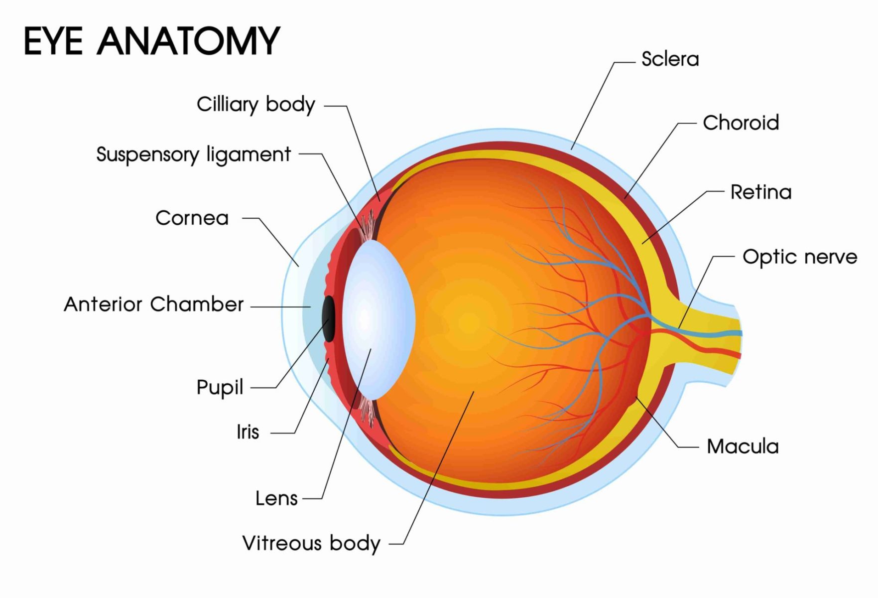
Guide To Eye Anatomy Diagram And Parts Of The Eye Explained Based on my experience here are three rules to live by: use the first pass to familiarize yourself with terminology. use the second pass to see how anatomical structures relate and fit together. use the third pass to memorize. when i say “passes”, i’m referring more to an initial glance at the materials here. The surface of the eye and the inner surface of the eyelids are covered with a clear membrane called the conjunctiva. the layers of the tear film keep the front of the eye lubricated. tears lubricate the eye and are made up of three layers. these three layers together are called the tear film. the mucous layer is made by the conjunctiva. Light is focused primarily by the cornea – the clear front surface of the eye, which acts like a camera lens. the iris (colored part) of the eye functions like the diaphragm of a camera, controlling the amount of light reaching the retina by automatically adjusting the size of the pupil (aperture). the eye’s crystalline lens is located. Understanding human anatomy is crucial for success in both education and healthcare. that’s why over 12 million students, educators, and professionals turn to teachmeanatomy for in depth guides, high quality visuals, interactive tools & study tips to help you understand the most complex anatomical concepts.

Page 14 Eye Anatomy Anatomy Science Light is focused primarily by the cornea – the clear front surface of the eye, which acts like a camera lens. the iris (colored part) of the eye functions like the diaphragm of a camera, controlling the amount of light reaching the retina by automatically adjusting the size of the pupil (aperture). the eye’s crystalline lens is located. Understanding human anatomy is crucial for success in both education and healthcare. that’s why over 12 million students, educators, and professionals turn to teachmeanatomy for in depth guides, high quality visuals, interactive tools & study tips to help you understand the most complex anatomical concepts. The eye is a complex structure with layers, lens, muscles, receptors, that is surrounded by many bones. i keep things simple in this video, and correlate directly with the anatomy chapter from the book. i’ve also scanned in an entire head ct to help you correlate the cartoons with real clinical imaging. here are screen captures from this video:. Your eye structure lets light enter and pass through a series of clear components and sections, including the cornea, aqueous humor, lens and vitreous humor. those structures bend and focus light, adjusting how far the light beams travel before they come into focus. the focus needs to be precise.

Comments are closed.